
A Deadly Pathogen Decimated Sunflower Sea Stars. Look Inside the Lab Working to Bring Them Back by Freezing and Thawing Their Larvae
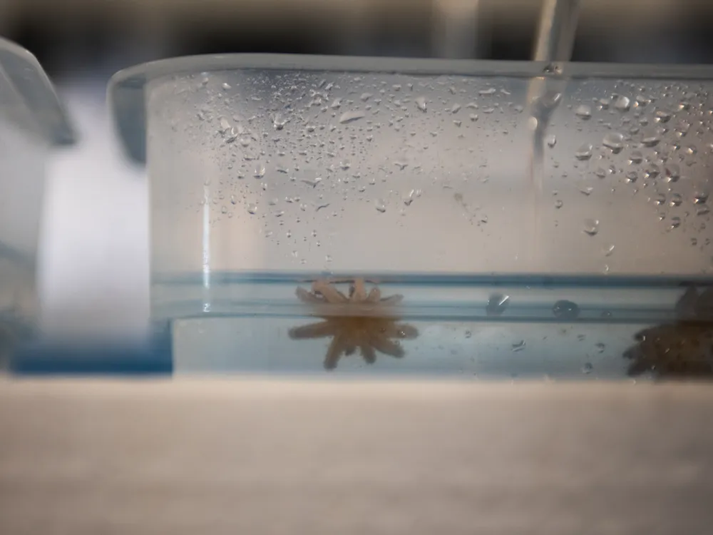
A Deadly Pathogen Decimated Sunflower Sea Stars. Look Inside the Lab Working to Bring Them Back by Freezing and Thawing Their Larvae For the first time, scientists have cryopreserved and revived the larvae of a sea star species. The breakthrough, made with the giant pink star, gives hope the technique could be repeated to save the imperiled predator Juvenile sunflower sea stars at the Sunflower Star Laboratory in Moss Landing, California. At this phase, each is less than an inch wide, but they can grow to be more than three feet across as adults. Avery Schuyler Nunn Key takeaways: Recovering sunflower sea stars by freezing them in time Ravaged by infectious bacteria, sunflower sea stars literally wasted away across the Pacific coast of North America—and their resulting population crash destabilized kelp forest ecosystems. Scientists pioneered a cryopreservation technique on the closely related giant pink star, raising hopes that a bank of frozen sunflower star larvae could one day be thawed in the same way and released into the wild. Along a working California harbor, where gulls wheel over weathered pilings and the old Western Flyer—the ship John Steinbeck once sailed to the Sea of Cortez—sits restored in its berth, researchers buzz about in a modest lab tucked between warehouses and boatyards. Inside, amid the hiss of pumps and the faint smell of brine from seawater tables, a scientist lifts a small vial from a plume of liquid nitrogen, its frosted casing holding the tiniest flicker of hope for a species on the brink. Each of the 18 vials contains between 500 and 700 larval giant pink sea stars. At this stage, they are tiny specks suspended in seawater, invisible to the naked eye. These particular larvae have been cryopreserved and stored at roughly minus 180 degrees Celsius since March. At the Sunflower Star Laboratory (SSL) in Moss Landing, California, scientists thawed the larval pink sea stars and coaxed them to successfully develop into juveniles this summer—a first for any sea star species. In October, the scientists thawed another batch of larvae from the same cohort to test larval growth and survival under different freezing conditions and thawing protocols. The breakthrough, however, isn’t really about the giant pink star, a species that’s common in the wild. Instead, these larvae serve as a crucial stand-in for the far more imperiled sunflower sea star (Pycnopodia helianthoides)—a vanishing species for which larvae are precious, limited and increasingly difficult to obtain. Perfecting cryopreservation methods on pink stars—ensuring they can survive freezing, resume feeding and grow into juveniles—lays the scientific groundwork for facilitating a return of Pycnopodia. The contents of a thawed vial are placed under a microscope to assess viability of the larvae. Avery Schuyler Nunn The discovery arrives at a precarious time, as sunflower stars have disappeared at a pace rarely seen in marine ecosystems. As a mysterious pathogen ravaged their population along the western shores of North America beginning in 2013, the creatures collapsed from an estimated six billion individuals to functional extinction in parts of their range—all within just a few years. Their loss left kelp forests with dramatically fewer predators, destabilizing ecosystems across the Pacific coast and allowing urchins to proliferate and graze formerly lush underwater canopies into barren rock. Now, scientists hope that “freezing” their larvae will offer a new avenue for bringing the species back. “Cryopreservation is particularly important on the population level when thinking about recovery for this endangered species, because it had major population losses,” says Marissa Baskett, an environmental scientist at the University of California, Davis, who was not involved in the project. The process lets scientists preserve the sea stars’ existing genetic diversity for future reintroduction to the wild, she adds. “Especially given the uncertainty about different disease outbreaks, having that stock to return to is incredibly valuable.” A mysterious and “complete collapse” Sunflower sea stars have long lived in abundance up and down the rugged Pacific coast—from Alaskan archipelagoes to Baja California. The 24-limbed echinoderms sprawled across the seafloor in shades of ochre, crimson and violet. Among the fastest-moving and largest of all sea stars—capable of stretching nearly three feet across—these radiant predators coursed through kelp forests, voraciously hunting purple sea urchins and preventing them from over-grazing on the holdfasts that root towering golden canopies of kelp. An adult sunflower sea star has 24 limbs and can be more than three feet wide. This one was photographed off Point Dume State Beach near Los Angeles. Brent Durand via Getty Images “In Northern California and Oregon, there historically would have been multiple keystone predators within the kelp forest ecosystem who are punching on purple urchins and keeping their population in check,” says Reuven Bank, board chair of SSL. “But the southern sea otter was extirpated across its historic range, so we were left with sunflower stars being the last major keystone predator of purple urchins across over 100 miles of coastline.” “And sunflower stars didn’t just eat urchins, they scared them,” Bank adds. “Urchins can smell a sunflower star approaching, and in healthy kelp forests they hide more and graze less. Even without consuming them, sunflower stars helped keep urchin behavior, and therefore kelp forests, in balance.” Then, in June 2013, tidepool monitors along Washington’s Olympic Peninsula documented an unprecedented sight. The once-sturdy sea stars had turned soft, pale and contorted, their arms curling and detaching from their bodies. By late summer, the same mysterious affliction had surfaced in British Columbia, and it began sweeping both north and south with startling speed. The emerging epidemic, which caused the invertebrates to literally disintegrate, would soon be known as sea star wasting disease. An infamous marine heatwave—nicknamed “The Blob”—had settled over the Pacific by 2014, thrusting the coast into a fever. Ocean temperatures spiked, likely speeding up the disease progression in already stressed sea stars and leading to higher mortality. In the warm, stagnant water, infected sunflower stars dissolved at an eerily rapid pace, leaving behind ghost-white films of bacterial mass where the vibrant predators had been just days before. “You’d have apparently healthy stars basically melt away into puddles of goo within 48 hours,” says Andrew Kim, lab manager at SSL. “It happened so quickly, and I don’t think folks were prepared for the ensuing ecosystem shift. You don’t often expect diseases to come through and totally reshape ecosystem dynamics within such a short period. But that’s what we saw.” Without sunflower sea stars to keep those spiny purple urchins in check, the balance began to falter, setting the stage for an unprecedented chain reaction. Urchin populations skyrocketed, grazing on kelp without limits, and once-thriving underwater forests collapsed into barren rock. A dense group of purple sea urchins, which exploded in population after the sunflower sea stars disappeared, photographed near Mendocino Headlands State Park, north of San Francisco. Brent Durand via Getty Images In California, with 99 percent loss, sunflower sea stars are now considered functionally extinct. “Even though there may be a few remnant individuals left, they can no longer fulfill their historic role in the ecosystem,” Bank says. As sunflower stars unraveled in the wild, another species—its thick-armed cousin, the giant pink star—offered an unexpected foothold for hope. The pink stars share a nearly identical geographic range and life history with sunflower stars, and crucially, their larvae can be raised in aquaria. If scientists could learn to freeze and revive the pink star in its early life stages, they wondered, could that knowledge become a lifeline for the sunflower star? That’s where the small team in Moss Landing stepped in. Freezing sea stars for the future What these scientists did was something no one had ever pulled off with a sea star. Working with giant pink stars, researchers spawned adults at the Aquarium of the Pacific in Long Beach, California, fertilized their gametes to produce thousands of larvae, and shipped those microscopic bodies to the Frozen Zoo—a cryopreserved archive of creatures operated by the San Diego Zoo Wildlife Alliance. There, reproductive scientists plunged the larvae into liquid nitrogen, cooling them to extremely low temperatures and pausing their cells’ biological activity. The larvae, essentially frozen in time, were shielded from ice crystal damage with special cryoprotectant mixtures. Sunflower Star Laboratory researchers remove a vial of pink star larvae from an insulated cooler at around minus 180 degrees Celsius in preparation for thawing. Avery Schuyler Nunn After months in this suspended state, the larvae were sent to the Sunflower Star Laboratory where Carly Young, a San Diego Zoo Wildlife Alliance scientist who advances cryopreservation and reproductive-rescue tools, led the team in thawing the vials. She had fine-tuned the ideal way to keep the larvae alive as they returned to real-world temperatures, carefully testing more than 100 “recipes” with various warming rates, cryoprotectant dilutions and rehydration steps. The pink star larvae not only survived thawing, but have thus far lived all the way through metamorphosis into juveniles. Scientists watched the little stars settle spontaneously along the bottom of their beakers just 19 days after revival. The success prompted the team to apply the same cryopreservation protocols to sunflower star larvae from the Alaska SeaLife Center. The larvae will be frozen in perpetuity, creating the first-ever cryopreserved archive of the species—like a seed bank, but for the baby sea stars. “A famous quote from the ’70s, when the Frozen Zoo in San Diego was established, was, ‘You must collect things for reasons you don’t yet understand,’” says Ashley Kidd, conservation project manager at SSL. “We don’t know when the other shoe is going to drop and what populations are going to look like as the planet changes. So, rather than chasing ghosts around the ocean floor, we really focused on what we can do with animals that are currently under human care somewhere.” While cryopreservation itself isn’t a ready-made restoration tool, it opens the door to conserving genetic diversity of a species and banking rare lineages for potential reintroduction to the wild. In the 1970s and 1990s, researchers began testing cryopreservation of marine invertebrates with sperm and larvae, establishing the basic protocols that this team could apply to sea stars. The breakthrough doesn’t restore kelp forests by itself, but the SSL scientists note that cryopreservation creates something the conservation community has desperately needed: time. Time to hold onto genetic diversity, time to refine captive rearing and time to prepare for future reintroduction at scales big enough to matter. The ultimate test, the researchers say, will be translating the thawing process to sunflower sea stars. Carly Young, at the Sunflower Star Laboratory, looks for movement in the young sea stars. Avery Schuyler Nunn Just this summer, scientists uncovered a piece of the puzzle that had eluded them for more than a decade: the pathogen behind sea star wasting disease. In a four-year international effort, researchers traced the outbreak to a strain of the marine bacterium Vibrio pectenicida. When cultured and injected into healthy sea stars, it reproduced the telltale symptoms—softening arms, rapid disintegration and death within days. The finding, published in Nature Ecology and Evolution in August, gives recovery teams a way to test for the pathogen in labs and hatcheries, tighten quarantine measures and understand disease risks before returning captive-bred sea stars to the Pacific. “It’s massively important to know what to look for, and the fact that we are now able to test for this disease is going to be critical in advancing our ability to move forward with reintroductions and continuing the research,” notes Kim. “We’ve already been able to take fluid samples from all of our stars and get them analyzed for the presence of Vibrio pectenicida, so we’ve mobilized very quickly on the heels of development.” Paired with this new diagnostic clarity, advances in cryopreservation offer a second front in the effort to save the species. Frozen larvae can be stored for decades and offer flexibility for selective breeding of disease-tolerant traits, notes the team. Cryopreservation adds another tool to the scientists’ toolbox as they fight to prevent the species—and, in turn, its ecosystem—from wasting away. “Bringing back sunflower stars,” Bank says, “is the single-most important step we can take toward restoring kelp forest balance.” Get the latest Science stories in your inbox.
For the first time, scientists have cryopreserved and revived the larvae of a sea star species. The breakthrough, made with the giant pink star, gives hope the technique could be repeated to save the imperiled predator
A Deadly Pathogen Decimated Sunflower Sea Stars. Look Inside the Lab Working to Bring Them Back by Freezing and Thawing Their Larvae
For the first time, scientists have cryopreserved and revived the larvae of a sea star species. The breakthrough, made with the giant pink star, gives hope the technique could be repeated to save the imperiled predator
Key takeaways: Recovering sunflower sea stars by freezing them in time
- Ravaged by infectious bacteria, sunflower sea stars literally wasted away across the Pacific coast of North America—and their resulting population crash destabilized kelp forest ecosystems.
- Scientists pioneered a cryopreservation technique on the closely related giant pink star, raising hopes that a bank of frozen sunflower star larvae could one day be thawed in the same way and released into the wild.
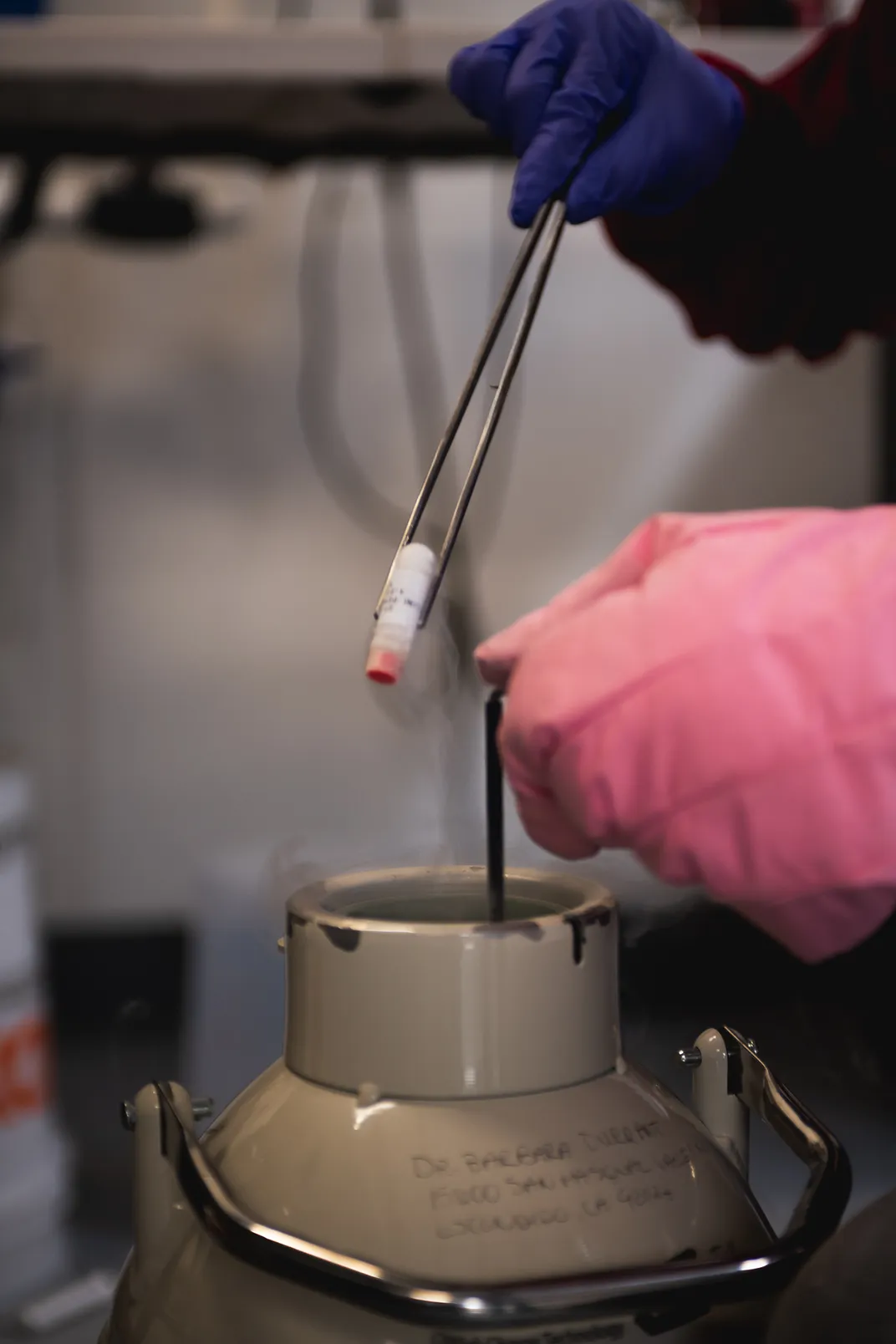
Along a working California harbor, where gulls wheel over weathered pilings and the old Western Flyer—the ship John Steinbeck once sailed to the Sea of Cortez—sits restored in its berth, researchers buzz about in a modest lab tucked between warehouses and boatyards. Inside, amid the hiss of pumps and the faint smell of brine from seawater tables, a scientist lifts a small vial from a plume of liquid nitrogen, its frosted casing holding the tiniest flicker of hope for a species on the brink.
Each of the 18 vials contains between 500 and 700 larval giant pink sea stars. At this stage, they are tiny specks suspended in seawater, invisible to the naked eye. These particular larvae have been cryopreserved and stored at roughly minus 180 degrees Celsius since March.
At the Sunflower Star Laboratory (SSL) in Moss Landing, California, scientists thawed the larval pink sea stars and coaxed them to successfully develop into juveniles this summer—a first for any sea star species. In October, the scientists thawed another batch of larvae from the same cohort to test larval growth and survival under different freezing conditions and thawing protocols.
The breakthrough, however, isn’t really about the giant pink star, a species that’s common in the wild. Instead, these larvae serve as a crucial stand-in for the far more imperiled sunflower sea star (Pycnopodia helianthoides)—a vanishing species for which larvae are precious, limited and increasingly difficult to obtain. Perfecting cryopreservation methods on pink stars—ensuring they can survive freezing, resume feeding and grow into juveniles—lays the scientific groundwork for facilitating a return of Pycnopodia.

The discovery arrives at a precarious time, as sunflower stars have disappeared at a pace rarely seen in marine ecosystems. As a mysterious pathogen ravaged their population along the western shores of North America beginning in 2013, the creatures collapsed from an estimated six billion individuals to functional extinction in parts of their range—all within just a few years. Their loss left kelp forests with dramatically fewer predators, destabilizing ecosystems across the Pacific coast and allowing urchins to proliferate and graze formerly lush underwater canopies into barren rock. Now, scientists hope that “freezing” their larvae will offer a new avenue for bringing the species back.
“Cryopreservation is particularly important on the population level when thinking about recovery for this endangered species, because it had major population losses,” says Marissa Baskett, an environmental scientist at the University of California, Davis, who was not involved in the project. The process lets scientists preserve the sea stars’ existing genetic diversity for future reintroduction to the wild, she adds. “Especially given the uncertainty about different disease outbreaks, having that stock to return to is incredibly valuable.”
A mysterious and “complete collapse”
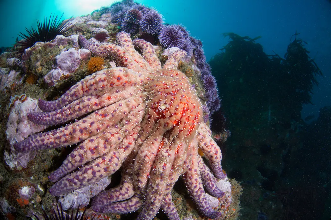
Sunflower sea stars have long lived in abundance up and down the rugged Pacific coast—from Alaskan archipelagoes to Baja California. The 24-limbed echinoderms sprawled across the seafloor in shades of ochre, crimson and violet. Among the fastest-moving and largest of all sea stars—capable of stretching nearly three feet across—these radiant predators coursed through kelp forests, voraciously hunting purple sea urchins and preventing them from over-grazing on the holdfasts that root towering golden canopies of kelp.
“In Northern California and Oregon, there historically would have been multiple keystone predators within the kelp forest ecosystem who are punching on purple urchins and keeping their population in check,” says Reuven Bank, board chair of SSL. “But the southern sea otter was extirpated across its historic range, so we were left with sunflower stars being the last major keystone predator of purple urchins across over 100 miles of coastline.”
“And sunflower stars didn’t just eat urchins, they scared them,” Bank adds. “Urchins can smell a sunflower star approaching, and in healthy kelp forests they hide more and graze less. Even without consuming them, sunflower stars helped keep urchin behavior, and therefore kelp forests, in balance.”
Then, in June 2013, tidepool monitors along Washington’s Olympic Peninsula documented an unprecedented sight. The once-sturdy sea stars had turned soft, pale and contorted, their arms curling and detaching from their bodies. By late summer, the same mysterious affliction had surfaced in British Columbia, and it began sweeping both north and south with startling speed. The emerging epidemic, which caused the invertebrates to literally disintegrate, would soon be known as sea star wasting disease.
An infamous marine heatwave—nicknamed “The Blob”—had settled over the Pacific by 2014, thrusting the coast into a fever. Ocean temperatures spiked, likely speeding up the disease progression in already stressed sea stars and leading to higher mortality. In the warm, stagnant water, infected sunflower stars dissolved at an eerily rapid pace, leaving behind ghost-white films of bacterial mass where the vibrant predators had been just days before.
“You’d have apparently healthy stars basically melt away into puddles of goo within 48 hours,” says Andrew Kim, lab manager at SSL. “It happened so quickly, and I don’t think folks were prepared for the ensuing ecosystem shift. You don’t often expect diseases to come through and totally reshape ecosystem dynamics within such a short period. But that’s what we saw.”
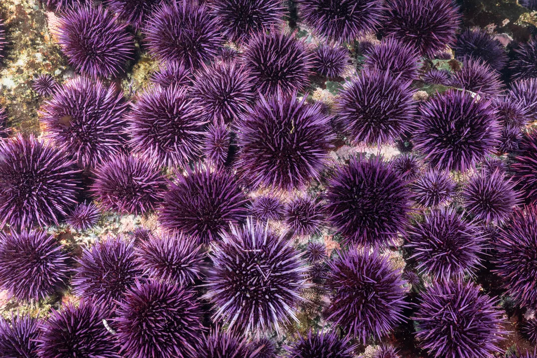
Without sunflower sea stars to keep those spiny purple urchins in check, the balance began to falter, setting the stage for an unprecedented chain reaction. Urchin populations skyrocketed, grazing on kelp without limits, and once-thriving underwater forests collapsed into barren rock.
In California, with 99 percent loss, sunflower sea stars are now considered functionally extinct. “Even though there may be a few remnant individuals left, they can no longer fulfill their historic role in the ecosystem,” Bank says.
As sunflower stars unraveled in the wild, another species—its thick-armed cousin, the giant pink star—offered an unexpected foothold for hope. The pink stars share a nearly identical geographic range and life history with sunflower stars, and crucially, their larvae can be raised in aquaria. If scientists could learn to freeze and revive the pink star in its early life stages, they wondered, could that knowledge become a lifeline for the sunflower star?
That’s where the small team in Moss Landing stepped in.
Freezing sea stars for the future
What these scientists did was something no one had ever pulled off with a sea star. Working with giant pink stars, researchers spawned adults at the Aquarium of the Pacific in Long Beach, California, fertilized their gametes to produce thousands of larvae, and shipped those microscopic bodies to the Frozen Zoo—a cryopreserved archive of creatures operated by the San Diego Zoo Wildlife Alliance. There, reproductive scientists plunged the larvae into liquid nitrogen, cooling them to extremely low temperatures and pausing their cells’ biological activity. The larvae, essentially frozen in time, were shielded from ice crystal damage with special cryoprotectant mixtures.
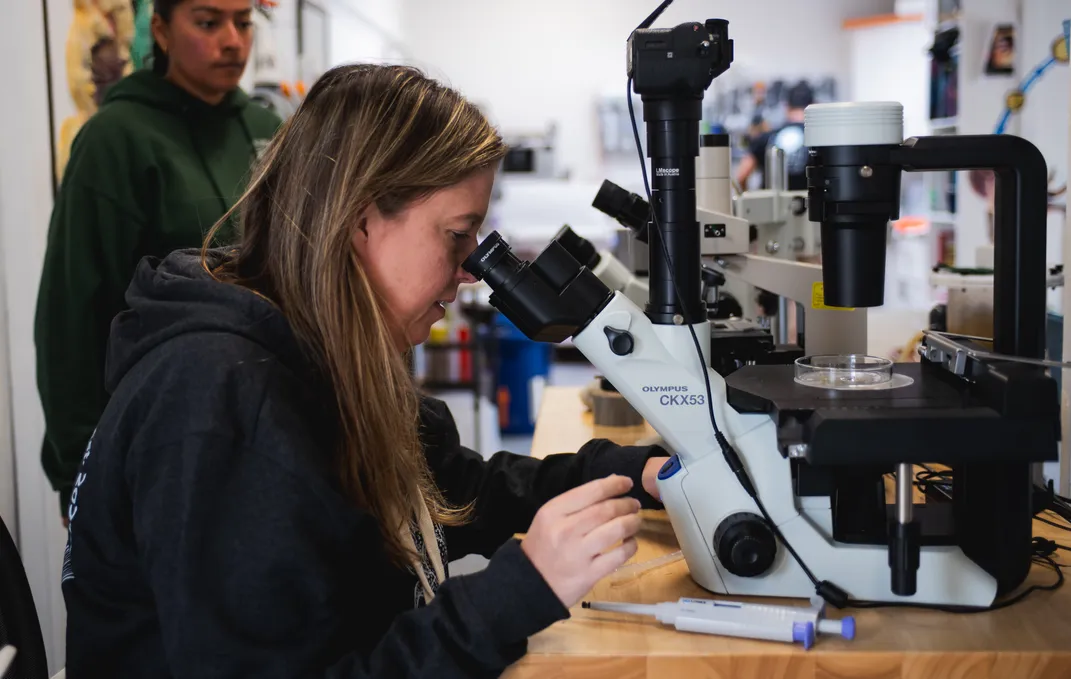
After months in this suspended state, the larvae were sent to the Sunflower Star Laboratory where Carly Young, a San Diego Zoo Wildlife Alliance scientist who advances cryopreservation and reproductive-rescue tools, led the team in thawing the vials. She had fine-tuned the ideal way to keep the larvae alive as they returned to real-world temperatures, carefully testing more than 100 “recipes” with various warming rates, cryoprotectant dilutions and rehydration steps.
The pink star larvae not only survived thawing, but have thus far lived all the way through metamorphosis into juveniles. Scientists watched the little stars settle spontaneously along the bottom of their beakers just 19 days after revival.
The success prompted the team to apply the same cryopreservation protocols to sunflower star larvae from the Alaska SeaLife Center. The larvae will be frozen in perpetuity, creating the first-ever cryopreserved archive of the species—like a seed bank, but for the baby sea stars.
“A famous quote from the ’70s, when the Frozen Zoo in San Diego was established, was, ‘You must collect things for reasons you don’t yet understand,’” says Ashley Kidd, conservation project manager at SSL. “We don’t know when the other shoe is going to drop and what populations are going to look like as the planet changes. So, rather than chasing ghosts around the ocean floor, we really focused on what we can do with animals that are currently under human care somewhere.”
While cryopreservation itself isn’t a ready-made restoration tool, it opens the door to conserving genetic diversity of a species and banking rare lineages for potential reintroduction to the wild. In the 1970s and 1990s, researchers began testing cryopreservation of marine invertebrates with sperm and larvae, establishing the basic protocols that this team could apply to sea stars.
The breakthrough doesn’t restore kelp forests by itself, but the SSL scientists note that cryopreservation creates something the conservation community has desperately needed: time. Time to hold onto genetic diversity, time to refine captive rearing and time to prepare for future reintroduction at scales big enough to matter. The ultimate test, the researchers say, will be translating the thawing process to sunflower sea stars.
Just this summer, scientists uncovered a piece of the puzzle that had eluded them for more than a decade: the pathogen behind sea star wasting disease. In a four-year international effort, researchers traced the outbreak to a strain of the marine bacterium Vibrio pectenicida. When cultured and injected into healthy sea stars, it reproduced the telltale symptoms—softening arms, rapid disintegration and death within days. The finding, published in Nature Ecology and Evolution in August, gives recovery teams a way to test for the pathogen in labs and hatcheries, tighten quarantine measures and understand disease risks before returning captive-bred sea stars to the Pacific.
“It’s massively important to know what to look for, and the fact that we are now able to test for this disease is going to be critical in advancing our ability to move forward with reintroductions and continuing the research,” notes Kim. “We’ve already been able to take fluid samples from all of our stars and get them analyzed for the presence of Vibrio pectenicida, so we’ve mobilized very quickly on the heels of development.”
Paired with this new diagnostic clarity, advances in cryopreservation offer a second front in the effort to save the species. Frozen larvae can be stored for decades and offer flexibility for selective breeding of disease-tolerant traits, notes the team. Cryopreservation adds another tool to the scientists’ toolbox as they fight to prevent the species—and, in turn, its ecosystem—from wasting away.
“Bringing back sunflower stars,” Bank says, “is the single-most important step we can take toward restoring kelp forest balance.”